Figure 1.
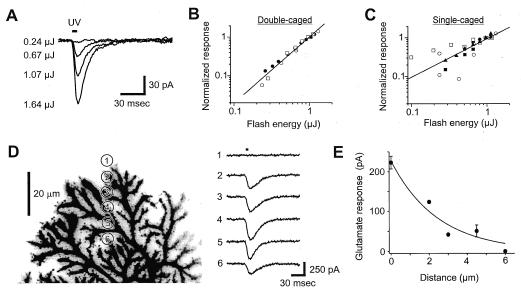
Nonlinear and localized activation of glutamate receptors by chemical two-photon uncaging. (A) Current responses to single-caged glutamate photolyzed by 6-ms UV flashes (at bar) of varying intensities focused on the soma of a Purkinje neuron. (B and C) Relationships between peak inward currents and light flash energy for double-caged (B) and single-caged (C) glutamate. Responses are normalized to the responses measured at 1 μJ. Each symbol represents a different cell. Least-squares fits to the data are shown by lines, with slopes of 1.7 in B and 1.0 in C. (D Left) Glutamate was produced by moving the uncaging spot to the locations indicated by numbered circles. This Purkinje cell was filled with Oregon Green 488 BAPTA-1 and contrast on this and all other fluorescence images in this paper is reversed, so that fluorescence is dark. (D Right) Responses elicited by uncaging glutamate (bar) at the locations shown (left). (E) Relationship between glutamate response and position of the UV light spot beyond the end of a Purkinje cell dendrite. Error bars indicate the SEM for points that represent the means of several measurements (n = 14 for 0 μm; n = 2 for 4.5 μm). Curve is an exponentially decaying function with a length constant of 2.4 μm.

