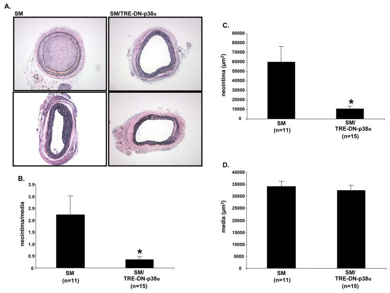
Figure 2. SM/TRE-DN-p38α compound transgenic mice exhibit reduced carotid injury-induced neointimal lesion formation.
A, SM and SM/TRE-DN-p38α mice were given doxycycline and mechanical injury of the left carotid artery was performed. Three weeks post-mechanical injury, histological examination with hematoxylin and eosin, and Verhoeff’s von Giesen stain was performed to assess neointimal lesion formation. Representative sections are depicted for SM and SM/TRE-DN-p38α mice; n=2 mice per genotype. A 10x objective was used to collect the micrographs. B, Mechanical injury and histology was performed as described for “A”. Then, morphometry was performed to quantify neointima/media ratios (SM/TRE-DN-p38α mice (n=15), + doxycycline vs. SM mice (n=11), + doxycycline, *P=0.029). C, Neointimal area was quantified via morphometric analysis with the mice from “A” and “B” (SM/TRE-DN-p38α mice (n=15), + doxycycline vs. SM mice (n=11), + doxycycline, *P=0.017). D, Medial area was quantified via morphometric analysis with the mice from “A” and “B”. For the bar graphs in “B”, “C”, and “D”, results are expressed as mean ± SE.

