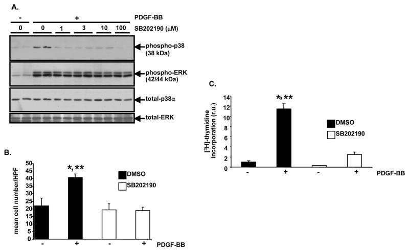
Figure 4. SB202190 blocks PDGF-induced p38 MAPK activation, cell proliferation, and DNA replication in A10 cells.
A, Analysis of p38 MAPK activation after SB202190 treatment. Serum-deprived A10 cells were treated with the indicated concentrations of SB202190 for 1 hour. Then, they were treated with or without PDGF-BB (60 ng/ml) for 5 minutes at 37 degrees C. Following stimulation, lysates were generated and Western blotting with anti-phospho (Thr180/Tyr182) p38 MAPK antibody was conducted. Subsequent re-blotting was conducted with anti-phospho (Thr202/Tyr204) ERK, anti-total p38α MAPK, and anti-total ERK antibodies. B, Evaluation of PDGF-stimulated cell proliferation after SB202190 treatment. Serum-deprived A10 cells were pre-treated with SB202190 (10 μM) or vehicle (DMSO) for 1 hour. Then, in triplicate wells, the monolayers were treated with PDGF-BB (60 ng/ml) or 0.1% BSA, as a vehicle control for PDGF-BB, in the continued presence of SB202190 or DMSO. After 20 hours cell number was determined. HPF, 400x high power field (DMSO, + PDGF-BB vs. DMSO, −PDGF-BB, *P<0.05; DMSO, + PDGF-BB vs. SB202190, − PDGF-BB, **P<0.05). C, Examination of PDGF-induced DNA replication after SB202190 treatment. Serum-deprived A10 cells were pre-treated with SB202190 (10 μM) or DMSO as in “B”. Then, the monolayers were treated with PDGF-BB (60 ng/ml) or 0.1% BSA for 15 hours in a tissue culture incubator in the continued presence of SB202190 or DMSO. Next, each well was pulsed with [6-3H]-thymidine (1 μCi/ml) for 2.5 hours and thymidine incorporation was determined; r.u., relative units (DMSO, + PDGF-BB vs. DMSO, −PDGF-BB, *P<0.05; DMSO, + PDGF-BB vs. SB202190, + PDGF-BB, **P<0.05). For bar graphs in “B” and “C”, results are expressed as mean ± SD of experiments conducted in triplicate.

