
Figure 3.
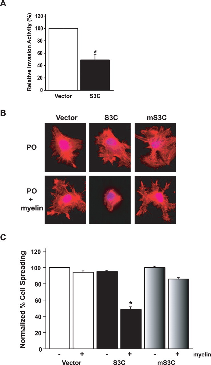
STAT3 decreases glioblastoma cell invasiveness and spreading on myelin. A, Matrigel invasion assay of U87 glioblastoma stable cell lines. Significantly more U87-vector cells invaded the matrigel substrate than U87-S3C cells (n = 3; t test, *p < 0.05). STAT3 inhibition of U87 glioblastoma cell invasiveness was not secondary to the effect of activated STAT3 on U87 cell proliferation, because the invasive potential of these cells was measured at a time (22 h after plating) before the inhibitory effects of activated STAT3 on U87 cell proliferation (Fig. 2B). Equivalent numbers of NIH3T3 cells failed to invade the matrigel (data not shown). B, Phalloidin red staining of actin stress fibers of stable U87 glioblastoma cells plated onto coverslips coated with either polyornithine (PO) or polyornithine together with myelin (20 μg/ml). U87-S3C glioblastoma cells failed to spread on myelin (middle bottom panel), compared with a spread appearance on a polyornithine control substrate (middle top panel). U87-vector and U87-mS3C glioblastoma cells spread and formed stress fibers on myelin (right and left bottom panels). C, Quantification of cell spreading of U87 stable cells on myelin. Significantly fewer U87-S3C glioblastoma cells spread on myelin compared with U87-vector glioblastoma cells (n = 3; ANOVA, *p < 0.0001). Cells were counted in a blinded manner in three independent experiments, and the percentage of spreading was determined by calculating the number of spread cells over the total number of cells.