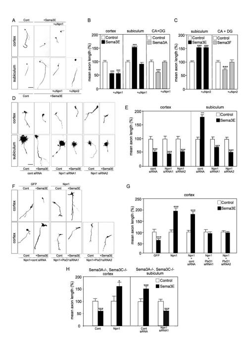
Fig. 6. Levels of Npn-1 gate the responses of cortical and subicular axons to Sema3E.
(A) Typical images of dissociated neurons cultured in the presence or absence of 10 μg/ml polyclonal anti-Npn-1 or anti-Npn-2, and of Sema3E. Blockade of Npn-1 prevents stimulation of subicular axon growth by Sema3E, but not inhibition of cortical axon growth by Sema3E. Anti-Npn-2 antibodies do not affect the growth-promoting effect of Sema3E on subicular axons. (B, C) Quantification of the results illustrated in A. Data are presented as mean axonal length ± s.e.m. (n=3) and are normalized to 100% for values obtained in control conditions. The anti-Npn-1 and anti-Npn-2 antibodies prevent inhibition of hippocampal axon growth by Sema3A (B) and Sema3F (C), respectively, confirming their neutralization efficiency. (D) Typical images of dissociated neurons cultured in the presence or absence of 5 nM Sema3E after electroporation of the indicated siRNAs. Knock-down of Npn-1 causes subicular neurons to switch their response to Sema3E from promotion to inhibition but does not affect inhibition of cortical axon growth by Sema3E. (E) Quantification of the results illustrated in D. Data are presented as mean axonal length ± s.e.m. (n= 3) and are normalized to 100% for values obtained in control conditions. (F) Typical images of dissociated neurons cultured in the presence or absence of 5 nM Sema3E after electroporation of expression vectors encoding GFP or Npn-1 together with the indicated siRNAs. Misexpression of Npn-1 confers on cortical neurons the ability to respond positively to Sema3E in a PlexinD1-dependent manner. (G) Quantification of the results illustrated in F. Data are presented as mean axonal length ± s.e.m. (n= 3) and are normalized to 100% for values obtained in control conditions. (H) To exclude the possibility that Npn-1 was acting as a receptor for other semaphorins produced in an autocrine manner (Bachelder et al., 2003; Serini et al., 2003; De Wit et al; 2005), responses of neurons from double mutant Sema3A-/-; Sema3C-/- embryos to Sema3E were quantified. Data are presented as mean axonal length ± s.e.m. (n= 3) and are normalized to 100% for values obtained in control conditions. Even in these mutant cells, misexpression of Npn-1 in cortical neurons and knock-down of Npn-1 in subicular neurons induce switches in axonal responses to Sema3E that are similar to those observed using wild-type neurons.
CA: cornus ammonis, DG: dentate gyrus. *significantly different with p<0.05; *** significantly different with p<0.001. Scale bar: 30 μm (A, D, F).

