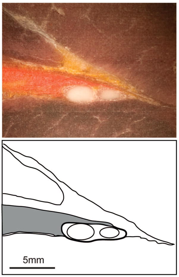
Fig. 2.
Photograph and reference tracing of a cryosection of the sciatic nerve and surrounding tissue after injection of 0.5 ml orange ink through a block needle that produced motor stimulation with a threshold of 0.3 mA in a control dog. The needle track does not appear in this section. The epineurium of the sciatic nerve is shown in the tracing with a heavy line, and the main fascicles within the nerve are shown by lighter lines. The ink distribution is represented in gray shading. The ink layers against the external aspect of the epineurium of the nerve, and the injection pattern was categorized as in contact with the nerve.

