Fig. 2.
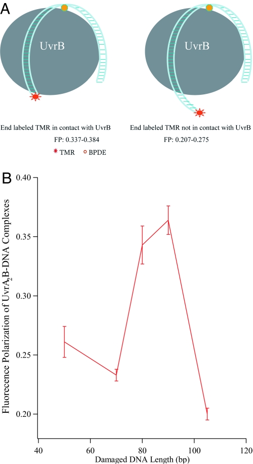
DNA wrapping of UvrB. (A) Schematic illustration of DNA wrapping of UvrB. Long DNA probes can be wrapped by UvrB, leading to multiple sites of contact between DNA and protein. The complexes would display large polarization responses if the end-labeling dye comes into close contact with the protein (Left), but only a moderate polarization value if the end-labeling TMR does not contact the protein because the DNA probe is too long (Right). Oligonucleotide duplexes that are too short to wrap around UvrB would not be expected to exhibit enhanced fluorescence polarization (not shown). (B) The fluorescence polarization of UvrAUvrB–DNA complexes as a function of damaged DNA length, confirming multipoint contact and its role in the large fluorescence-polarization response. The oligonucleotide sequences are shown in Table S2.

