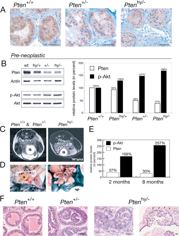
Figure 2. Ptenhy/− Mice Display Massive Hyperplasia and Invasive CaP.
(A) IHC on preneoplastic anterior prostate (AP) tissue of 2-mo-old littermate mice shows decreasing Pten protein levels in the hypomorphic series.
(B) WB analysis of the prostate lobes from (A) is shown on the left and their quantification is shown on the right.
(C) MRI of Ptenhy/− mice aged 6–8 mo shows pathologic structures (encircled by dashed lines) adjacent to seminal vesicles (SV) coinciding with displacement of the bladder (Bl), features typically associated with massive prostate tumors (right). These features were never found in Pten+/+ or Pten+/− mice (left).
(D) Macroscopic view of the Ptenhy/− mouse from (C) confirms massive enlargement of the AP lobes, but normal-sized seminal vesicles.
(E) Quantification of WB analysis on AP lobe total lysates from the Ptenhy/− animal shown in (C) compared to preneoplastic AP of same genotype from (A) and (B), labeled 8 mo and 2 mo, respectively. Note that in the enlarged hyperplastic prostate, Pten protein expression is retained (levels are expressed relative to the corresponding wild-type animals).
(F) Histopathology (H&E) of AP lesions at 8 mo reveals transition from low- to high-grade PIN and invasion (infiltration of stromal tissue is indicated by arrows) in Ptenhy/−, while age-matched Pten+/− tissue only shows hyperplastic features and Pten+/+ tissue is unaffected. Bars are 50 μm.

