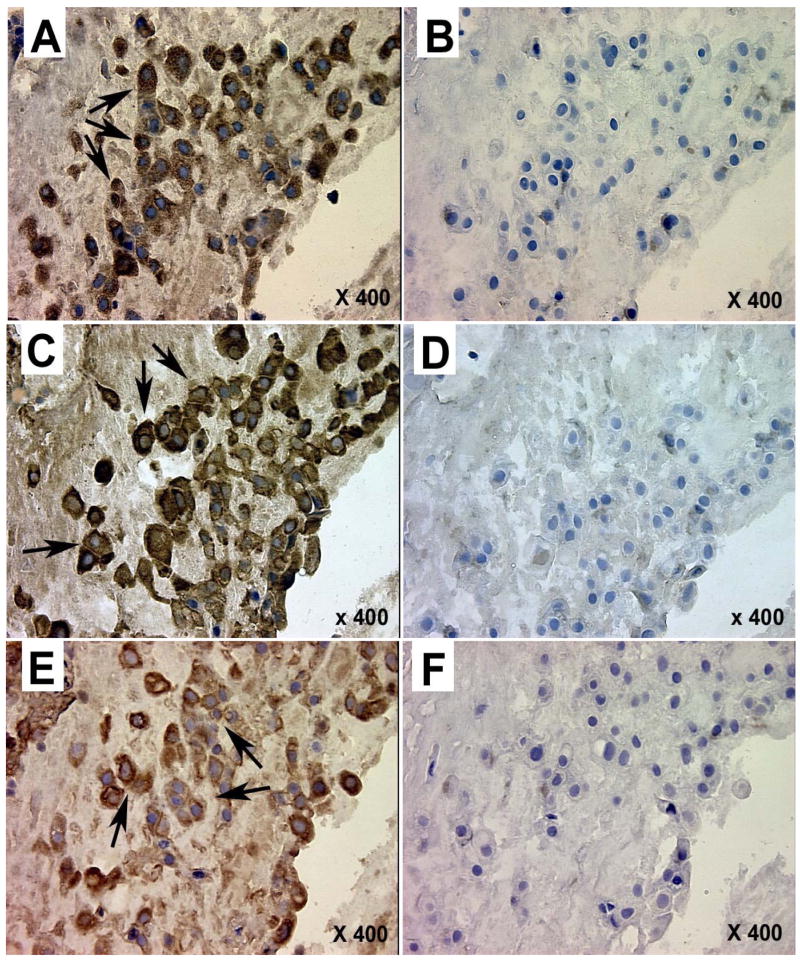
Figure 4.
Photomicrographs illustrating tissue localization of glutamate dehydrogenase (A, B), glutamine synthetase (C, D), and glutaminase (E, F) in sequential sections of the same placental fragment from a normal pregnancy. A, C, E – staining with primary antisera, B, D, F – normal rabbit serum controls. Arrows mark positive staining for cytotrophoblast cells (Langhans cells) grouped in columns within stem villi near the chorionic plate.

