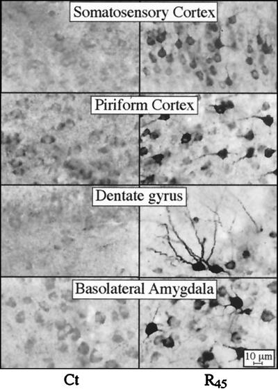
Figure 2.
AChE-R immunoreactivity in transgenic brain. Shown is AChE-R immunoreactivity in high-magnification photomicrographs of coronal sections 1.2 mm posterior to bregma from the brains of control (Ct) mice and the AChE-R line 45 transgenics (R45). Note that ARP immunostaining is apparent only in neurons, that both somata and processes are stained, and that the intensity of neuronal staining is highly variable between the different brain regions.

