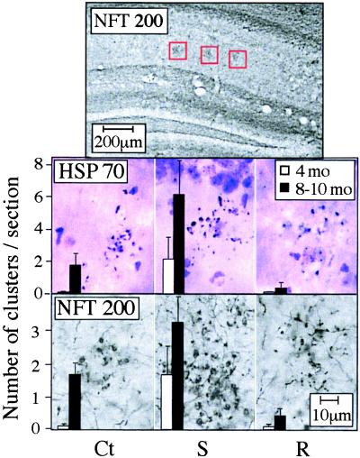
Figure 4.
Clustered neuronal fragments. Shown are coronal brain sections from 8- to 10-month-old control and transgenic mice after immunostaining of NFT200 or HSP70 with cresyl violet counter staining. (Top) Low-magnification image, in which gray-black clusters of neuronal fragments immunopositive for NFT200 (surrounded by red frames) are apparent in the stratum radiatum layer of hippocampal CA1–3 regions (1.6–2.8 mm posterior to bregma). (Middle and Bottom) High-magnification images of the framed regions, stained for HSP70 and NFT200, respectively. Clusters including over 10 neuronal fragments were counted in three sections from each mouse, 2 to 4 mm posterior to bregma. Bars present counted clusters per section as average ± SEM for six mice from each strain and age group (4-month-old and 8- to 10-month-old). Ct, control FVB/N mouse; S, AChE-S transgenic mouse; R, AChE-R transgenic mouse.

