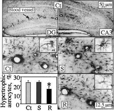
Figure 5.
Astrocyte reactivity. Normal and reactive astrocytes were counted in the stratum lacunosum moleculare in hippocampal sections 2 to 4 mm posterior to bregma (three from each of six mice in each strain). Shown is GFAP astrocyte labeling in coronal brain sections from 4-month-old transgenic and control mice. Note that in normal astrocytes, only dendrites are stained, and soma are pale or invisible, whereas reactive cells display highly immunoreactive soma and enhanced dendritic staining. (Top) Example low-magnification micrographs of dentate gyrus (DG; Left) and CA3 (Right) hippocampal regions of control (Ct) mice. (Middle and Bottom) High-magnification images of stratum lacunosum moleculare from the hippocampal CA3 subregion from control (Ct), AChE-S (S), and AChE-R (R) mice. (Insets) Individual astrocytes stained for GFAP (Bar = 1 μm). (Bottom Left) Percentage of reactive astrocytes of the total counted astrocytes (average ± SEM of n = 6 mice for each group). *, P < 0.05.

