1.

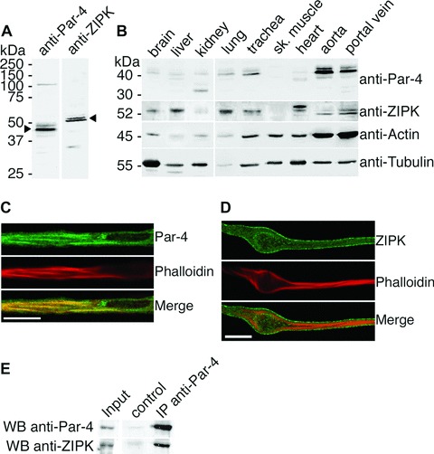
ZIPK and Par-4 protein expression, subcellular localization and in vivo interaction. (A) Full-length western blots demonstrate specificity of the Par-4 and ZIPK antibodies. Portal vein lysates are shown here. (B) Homogenates from ferret tissues (10 μg total protein per lane) subjected to western blot analysis with anti-Par-4 and anti-ZIPK antibodies. The same membrane was stained with anti-actin and anti-tubulin antibodies to assess equal protein loading and transfer. (C and D) Confocal immunofluorescence imaging of dVSMCs performed with (C) anti-Par-4 and (D) anti-ZIPK antibodies. Filamentous actin was stained with phalloidin. Scale bar, 10 μm. (E) Endogenous Par-4 was immunoprecipitated from A7r5 cell lysates performed with the monoclonal anti-Par-4 antibody. Co-immuno-precipitated endogenous ZIPK was detected by western blotting. For the control sample, A7r5 lysates were incubated with beads only.
