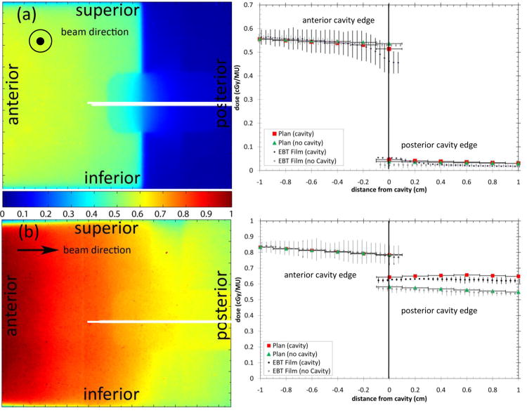
Figure 2.
Sagittal film results from (a) single laterally incident beam and (b) single anterior-posterior beam with and without a cavity. The white lines show the location of the profiles. The arrows show the beam direction. Horizontal error bars on the plan data show the width of the planned dose voxels.

