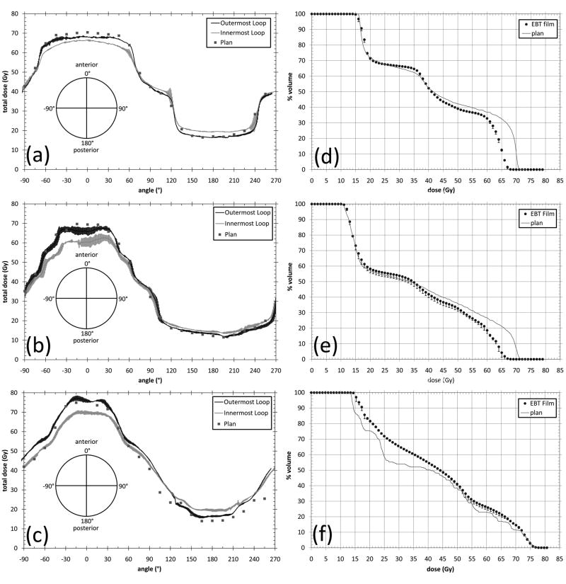
Figure 4.
Measured and planned rectal wall doses and resultant DVH from spiral film geometry. (a) represents the dose to the outermost and innermost loop of the film spiral and the planned dose to the film spiral for the 3DCRT plan (f) represents the resultant rectal wall DVH from the film spiral and the planned rectal wall DVH for the 3DCRT plan. (b) and (e), and (c) and (f) represent the same for the IMRT and helical tomotherapy plans respectively.

