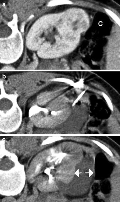
Fig. 1.
Prone contrast-enhanced CT images illustrating the technique of “hydrodissection.” a Image showing an exophytic renal tumor lying in close proximity to the colon (C). b A needle has been introduced into the perirenal space and 5% dextrose is being instilled. c The 5% dextrose has created a safety margin (arrows) between the tumor and adjacent colon. The tines of an expandable RFA probe can be seen within the lesion to be treated

