Fig. 3.
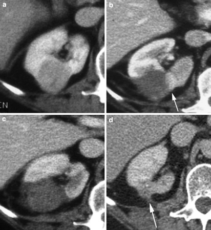
a Axial CT image of a 3-cm exophytic right interpolar renal tumor prior to RFA. b Ten days following RFA, contrast-enhanced CT reveals that the majority of the lesion is nonenhancing, consistent with necrosis, but there is a residual crescent of enhancing tumor within the medial aspect of the lesion (arrow). c One week after re-treatment of the residual crescent of tumor, the whole lesion is nonenhancing, in addition to a wedge-shaped area of adjacent cortex. This is consistent with complete necrosis of the tumor. d Five years post RFA: the lesion shows typical involution, with dispersal into the perirenal fat (arrow). There is no evidence of tumor recurrence

