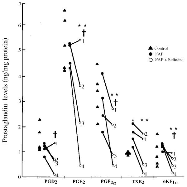
Fig 1.
Prostaglandin levels (ng/mg protein) in colorectal mucosa. PGD2, prostaglandin D2; PGE2, prostaglandin E2; PGF2α, prostaglandin F2α; TXB2, thromboxane B2; 6-keto-F1, 6-keto-prostaglandin F1α. FAP patients 1–4 are noted. * P = 0.016 control vs FAP; ** P < 0.05 FAP vs FAP and sulindac; † P < 0.05 control vs FAP and sulindac. Controls are five otherwise healthy Caucasians with normal colonoscopic examinations. FAP patients are four Caucasian patients with previous colectomy and ileorectal anastomosis. Patient 4 developed colorectal cancer after 35 months of sulindac treatment.

