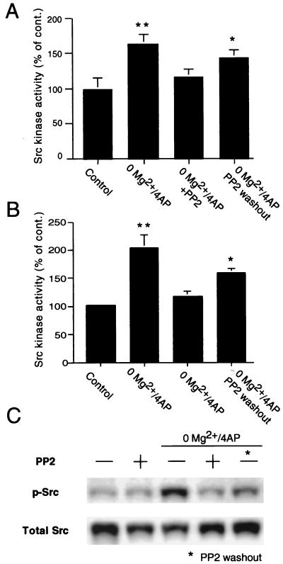
Figure 5.
Src kinase activity in extracts of hippocampal slices perfused with Mg2+/free medium containing 4-AP (50 μM) with or without PP2 (10 μM) and control slices. (A) Src kinase activity as measured by in vitro kinase assay in hippocampal extracts was significantly higher in slices perfused in Mg2+-free aCSF/4-AP (P < 0.01 from control slices incubated in normal aCSF). Incubation with PP2 prevented such an increase of Src activity (P < 0.01 from slices perfused in Mg2+-free aCSF/4-AP; P > 0.05 from control slices). One hour after PP2 washout, a partial recovery of Src kinase activity was seen. In fact, in these slices, Src activity was significantly higher than in control slices (P < 0.05), although not significantly different from slices treated with Mg2+-free aCSF/4-AP/PP2. (B) Similar results were obtained by quantification of catalytically active Src kinase in extracts of hippocampal slices by Western blot with an antibody specific for Src phosphorylated at the conserved residue Y418, which predicts kinase activation. Src phosphorylation was significantly induced in slices perfused in Mg2+-free aCSF/4-AP (P < 0.01 from control slices incubated in normal aCSF). Incubation with PP2 prevented such an increase of Src activity (P < 0.01 from slices perfused in Mg2+-free aCSF/4-AP; P > 0.05 from control slices). A recovery of Src phosphorylation was seen 1 h after PP2 washout (P > 0.05 from slices perfused in Mg2+-free aCSF/4-AP; P < 0.05 from control slices). (C) Representative Western blot. P-Src, phosphorylated Src (Y418); detection of total Src was used for control (total Src). With either method in the activity of Src kinase in slices treated with PP2 in normal aCSF did not differ from control slices in aCSF (not shown and C). *, Different from control P < 0.05; **, different from control P < 0.01.

