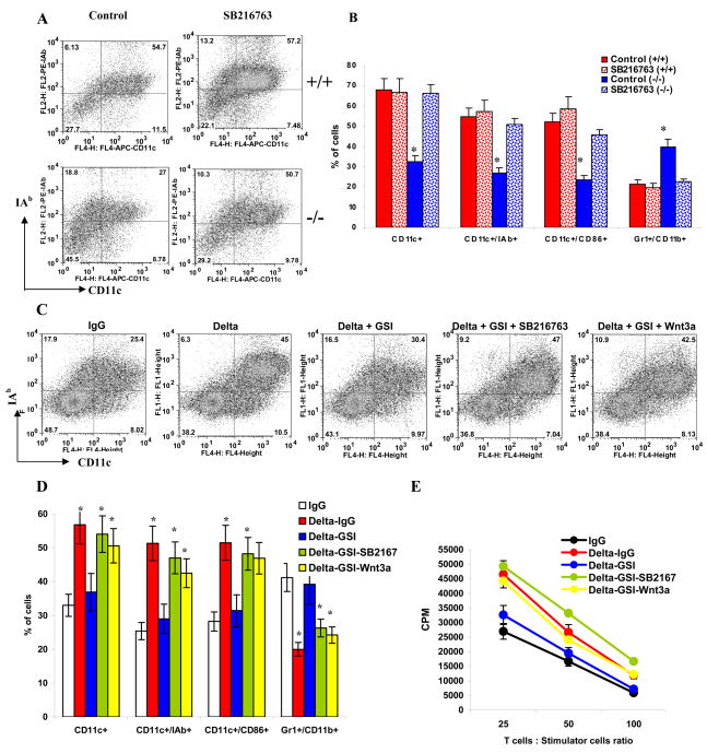
Figure 4. The role of Wnt signaling in Delta-1-mediated DC differentiation.
A, B DCs were differentiated from wild-type R1 ES cells and Notch-1−/− ES cells as described in Experimental Procedures. Cells were treated with 10 nM SB216763 or vehicle for 5 days prior to analysis. A. Typical example of one experiment. B. Cumulative results of three performed experiments. * - statistically significant (p<0.05) differences from control.
C,D. Enriched HPCs were cultured on plates coated with control IgG or Delta-IgG with or without GSI, SB216763, or Wnt3a as indicated. Cells were harvested and phenotype was evaluated by flow cytometry. C. Typical example of one experiment. D. Cumulative results of three performed experiments. * - statistically significant differences from IgG control (p<0.05). E. Allogeneic MLR. Cells were cultured with T cells isolated from allogeneic BALB/c mice for 4 days at different ratios. Cell proliferation was measured in triplicate by [3H]-thymidine uptake. Values are the mean ± SE.

