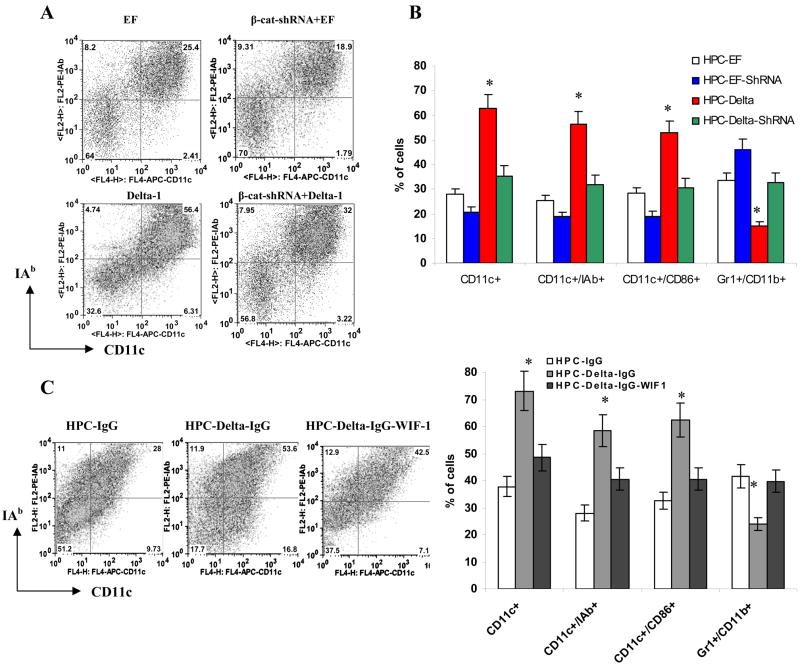
Figure 5. Cooperation between Notch and Wnt signaling in DC differentiation.
A, B. Enriched HPCs were transfected with β-catenin-shRNA-GFP construct or GFP vector, and then cultured on a monolayer of fibroblasts expressing Delta-1 or control vector (EF) for 5 days in CCM with 20 ng/ml GM-CSF. Cells were labeled with a cocktail of indicated antibodies and evaluated by flow cytometry. CD45+GFP+ cells were gated and the proportion of DCs or IMC was analyzed within this group of cells. A. Typical example of one experiment. B. Cumulative results of three performed experiments. * indicates statistically significant differences from EF control (p<0.05).
C. HPCs were incubated in CCM supplemented with 20 ng/ml GM-CSF for 5 days on a 24-well plate coated with Delta-1 protein or control IgG. Wif1 (750 ng/ml) was added at the start of the culture. Cell phenotype was evaluated by flow cytometry. Left – typical example of one experiment. Right - cumulative results of three performed experiments. * - statistically significant differences from IgG control (p<0.05).

