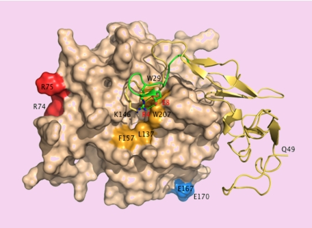
Surface representation of APC15 showing the back of the catalytic domain (wheat) connected to the EGF domains and the activation peptide (yellow ribbon). The A chain of thrombin9-comprising residues E1c through E8 (green ribbon) is superimposed for comparison. K146 (stick) occupies a position analogous to that of R4 (stick) in thrombin. Residue E149, not resolved in the crystal structure, could engage K146 in an ion-pair interaction as E8 (stick) does with R4 in thrombin. Disruption of the ion-pair with the E149A mutation could expose a hydrophobic patch (orange) for recognition of macromolecular ligands. Residues R74, R75, E167, and E170 (chymotrypsinogen numbering) face the front of the molecule and are only partially visible in this orientation. R74 and R75 are part of the factor Va epitope.6 E167 and E170 are involved in PAR1 recognition.7

An official website of the United States government
Here's how you know
Official websites use .gov
A
.gov website belongs to an official
government organization in the United States.
Secure .gov websites use HTTPS
A lock (
) or https:// means you've safely
connected to the .gov website. Share sensitive
information only on official, secure websites.
