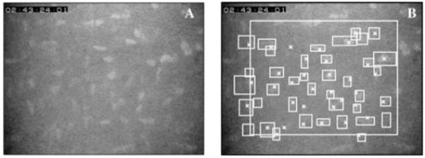
Fig. 2.

One confocal image before (A) and after counting cells (B). Cells were counted manually within or touching the left or bottom edge of the large rectangle and were indicated with an X. Cells identified by the program were indicated by a bounding box, the smallest rectangle with vertical and horizontal sides that entirely contained the cell. Most cells were identified by both methods, although some were identified by one or the other but not both.
