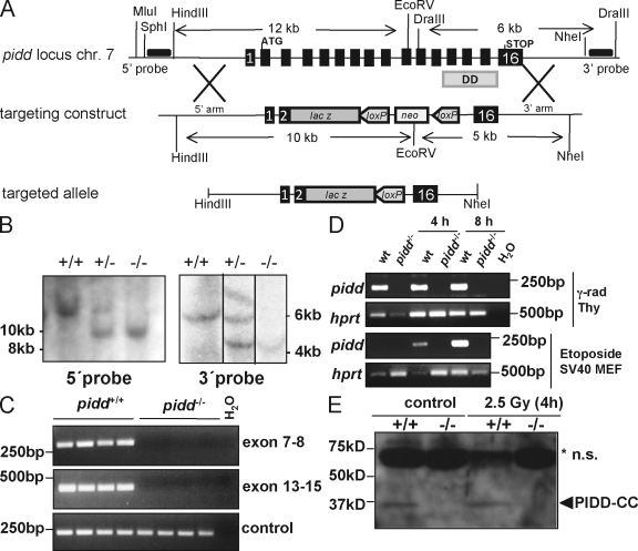
Figure 1.
Targeting the pidd locus in mice. (A) Schematic representation of the pidd/lrdd gene locus on mouse chromosome 7 (chr 7). Exons 3–15 of the pidd gene were replaced by the open reading frame of β-galactosidase. The loxP element-flanked neomycin selection marker cassette was removed in vivo by cre-mediated deletion. The locations of the 5′ and 3′ probes used for Southern blot analysis are indicated as horizontal black bars. (B) Southern blot analysis on genomic DNA derived from wild-type, pidd+/−, and pidd−/− mice. (C) Exon-specific PCR using three different primer pairs spanning exons 7–8 or 13–15 confirmed correct targeting of the pidd gene. (D) RT-PCR was performed on cDNA generated from total RNA derived from thymocytes (top two panels) that were left untreated or exposed to 2.5 Gy of γ irradiation or SV40-immortalized wild-type (wt) and PIDD-deficient MEFs (bottom two panels) cultured in the absence or presence of etoposide to confirm absence of pidd mRNA. (E) Western blot analysis on thymocyte extracts derived from wild-type and pidd−/− mice before and after exposure to γ irradiation in vitro confirmed absence of PIDD at the protein level. The asterisk indicates nonspecific (n.s.) bands.

