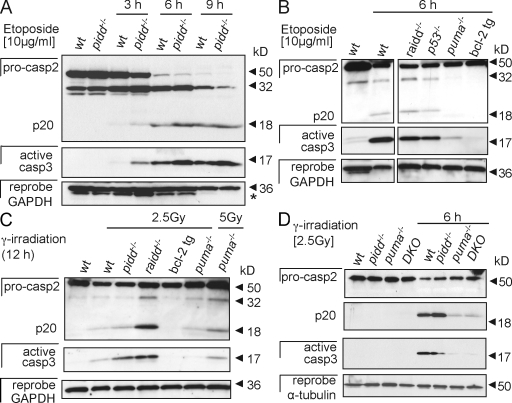
Figure 5.
Caspase-2 processing occurs in thymocytes lacking the PIDDosome. (A) Thymocytes isolated from wild-type (wt) and pidd−/− mice were cultured without further treatment or stimulated with etoposide. Caspase-2 processing and cleavage of effector caspase-3 was monitored over time by Western blot analysis. The asterisk indicates a residual caspase-2 signal remaining upon reprobing with anti-GAPDH antibody. (B and C) Caspase-2 and -3 processing in etoposide-treated (B) or γ-irradiated thymocytes (C) of the indicated genotypes. tg, transgenic. (D) Thymocytes isolated from wild-type, pidd−/−, puma−/−, and pidd−/−puma−/− mice were cultured without further treatment or after exposure to γ irradiation for 6 h. Caspase-2 processing and cleavage of effector caspase-3 were monitored by Western blot analysis. After detection of caspase-2, membranes were first stripped and reprobed with anti–active caspase-3 antibody and finally probed without stripping with an antibody recognizing GAPDH or α-tubulin to compare protein loading. DKO, double knockout.

