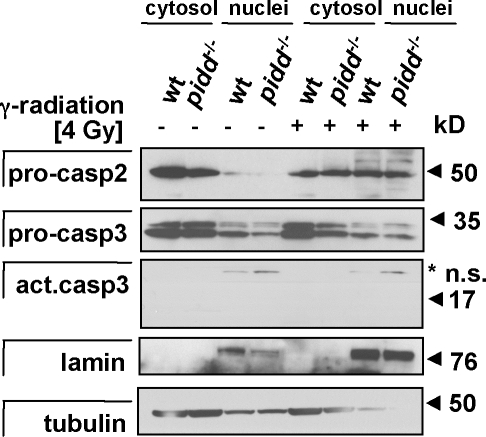
Figure 9.
Nuclear translocation of caspase-2 upon DNA damage. Cytosolic and nuclear extracts from wild-type (wt) and PIDD-deficient MEFs were generated from untreated cultures or 4 h after exposure to 4 Gy of γ irradiation. Extracts were separated by SDS-PAGE, and caspase-2 was detected by immunoblotting. Membranes were reprobed with antilamin- and antitubulin-specific antibodies to assess cross-contamination of fractions as well as anti–active caspase-3 to monitor for effector caspase activation. The asterisk indicates nonspecific (n.s.) bands.

