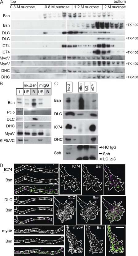
Figure 5.
DLC and other motor components associate with Bassoon transport packets. (A) Membranes of rat embryonic brain (E18) were subjected to a floatation assay. Equal volumes of each fraction were analyzed on WBs. Membrane association was tested by incubation with the detergent Triton X-100 (+TX-100), which prevents floating of membrane-associated proteins. (B) PTVs were isolated from the light membrane fraction (0.3–0.8 M sucrose) by immunoprecipitation with Bassoon-specific antibodies, solubilized, and probed for their protein content on WBs (mαBsn column). PTV marker proteins Bassoon (Bsn) and Piccolo (Pclo) were detected as well as DLC, DHC, and myosin V (myoV). KIF5A/C was not precipitated at detectable levels. Precipitation with unspecific IgGs (mIgG column) confirmed the specificity of binding. I, input; UB, unbound material; B, bound material. (C) Coprecipitation of protein complexes with anti-Bassoon (αBsn column) and anti-IC74 (αIC74 column) antibody from brain lysate of P1 rats. The presence of DLC in Bassoon-containing complexes as well as the association of Bassoon with assembled dynein motor complex (containing IC74, DHC, and DLC) can be seen. Lanes with input and control precipitations with unrelated IgG are indicated. Synaptophysin (Sph) was not detected in any of the immunoprecipitated complexes. HC, heavy chain; and LC, light chain of coupled antibody. (D) Costaining of Bassoon (magenta) with IC74, DLC1/2, and myosin V (green) in distal axons (left image sequences) and growth cones (right image sequences) of neurons at day 6 after plating. Outlines of axons and growth cones were created according to cell autofluorescences in raw images. Bar, 5 µm.

