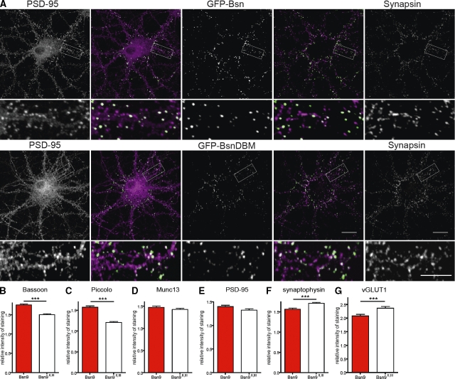
Figure 8.
Overexpression of dominant-negative fragment Bsn9 enhances accumulation of Bassoon and Piccolo at synapses. (A) Cells transfected at 3 DIV with GFP-Bsn (top) and GFP-BsnDBM (bottom) were fixed at 15 DIV and counterstained to visualize synapsin (last image in each row and magenta in merged image) and PSD-95 (first image in each row and magenta in merged image). The fluorescence of GFP fusion proteins is shown in the middle image of each row (green in merged images). The second and fourth panels are merged images of panels to their right and left. Higher magnifications of the boxed regions are shown below the respective image. (B–G) Quantification of relative staining intensities for Bassoon (B), Piccolo (C), Munc13 (D), PSD-95 (E), synaptophysin (F), and vGLUT1 (G) in cells transfected at 3 DIV with the bicistronic construct expressing EGFP-synapsin and Bsn9 (red bar) or control fragment Bsn9II,III (open bar) and analyzed at 9 DIV. Bar graphs show mean values of relative fluorescence intensity for each staining, and error bars indicate SEM. Values are derived from three independent experiments. ***, P < 0.001. Bars: (A) 20 µm; (insets) 10 µm.

