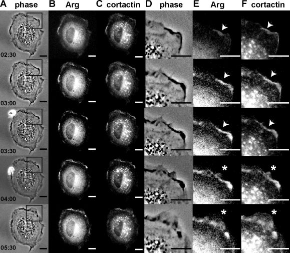
Figure 1.
Arg-YFP and cortactin-RFP colocalize to membrane protrusions in response to adhesion to fibronectin. Individual frames from time-lapse videos of arg−/− cells expressing Arg-YFP and cortactin-RFP (Video 1). (A–F) Phase-contrast (A and D), YFP channel (B and E), and RFP channel (C and F) images are shown. (D–F) Enlargements of the regions boxed in A, showing colocalization of Arg-YFP and cortactin-RFP (indicated by arrowheads) followed by the formation of a cell edge protrusion (indicated by asterisks). The elapsed time is shown in minutes:seconds. Bars, 10 µm.

