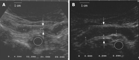
Figure 2.
A: Before infliximab therapy: A stenotic lesion of the terminal ileum, with narrow lumen (white arrows) and increased thickness of the intestinal wall. The markers assess the length. The iliac vessel is indicated (circle); B: After infliximab therapy (22 cycles): The same ileal segment with an increased lumen diameter (12 mm) and reduced thickness of the intestinal wall. The iliac vessel is indicated (circle).

