
Figure 3.
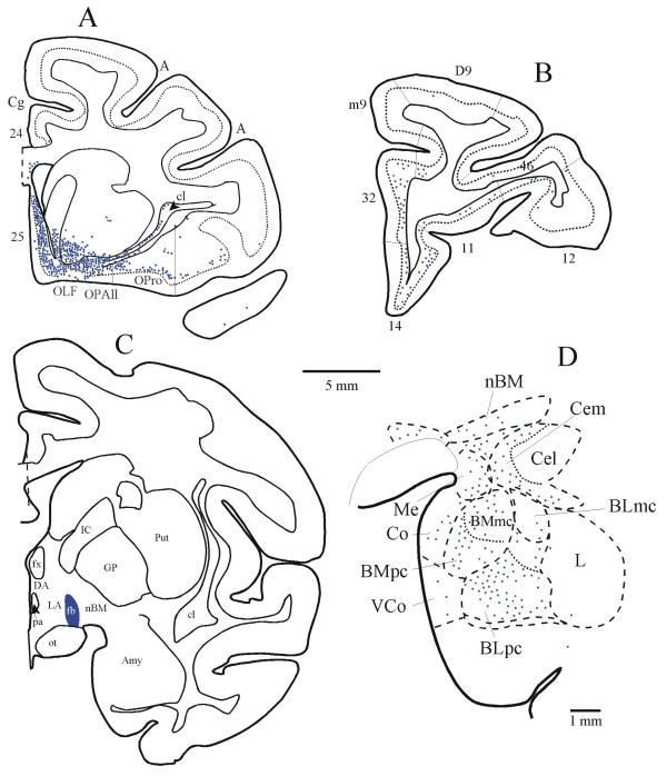
Simultaneous labelling of pathways from orbitofrontal cortex and the amygdala to the lateral hypothalamic area. (A) Densely distributed projection neurons in the posterior orbitofrontal cortex (areas OPAll, OPro), and in medial area 25 projected to the lateral hypothalamic area (LA in C). (B) Projection neurons in prefrontal areas 32 and 14 and in other prefrontal areas directed to LA; (C) The injection site of fast blue (fb) was in LA of the hypothalamus. (D) Projection neurons from the amygdala directed to the same area (LA) of the hypothalamus. The dotted line in A and B indicates the upper border of cortical layer V.