
Figure 2.
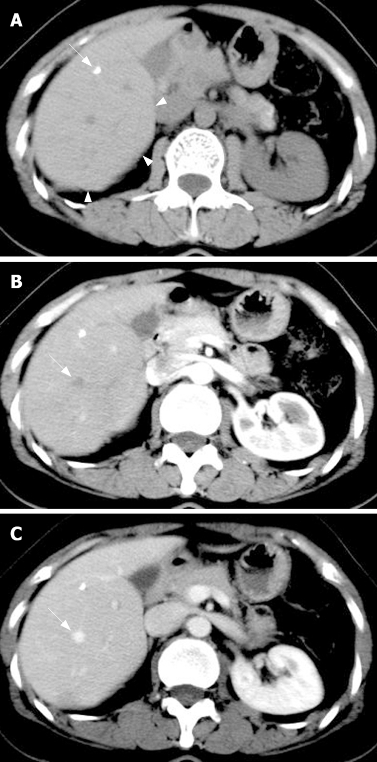
Transverse unenhanced CT image showing the protrudent visceral surface which hints a local iso-attenuated lesion (arrowheads) with punctate calcification (arrow) (A), identical density of the lesion to the adjacent liver parenchyma at arterial phase (B) and portal phase (C). The right hepatic vein with a normal shape and location crosses the lesion (arrow).