Figure 1.

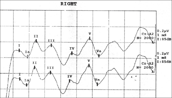
Brainstem auditory evoked potentials wave pattern over right ear of a healthy volunteer. Wave I and IPL of I-III represent the peripheral part of the pathway; whereas wave III and IPL of III-V, the central part

Brainstem auditory evoked potentials wave pattern over right ear of a healthy volunteer. Wave I and IPL of I-III represent the peripheral part of the pathway; whereas wave III and IPL of III-V, the central part