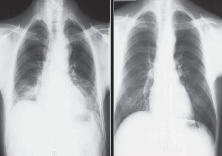
Figure 2.

Chest radiographs on the same patient a few minutes apart showing the effect of technique; the left image shows mediastinal widening and basal clouding due to a poor inspiratory effort; the right image has been taken in good inspiration and looks entirely normal
