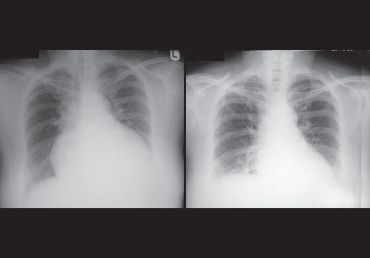
Figure 43.

The radiograph on the left was taken on admission of the patient, showing an enlarged globular heart secondary to pericardial effusion due to severe hypothyroidism. The image on the right was taken 3 weeks later, showing resolution of the pericardial effusion
