Figure 3.
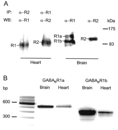
Detection of GABABR expressions and GABABR1 transcripts in heart and brain. (A) In whole-cell lysates of rat cardiomyocytes, AbC22 directed against GABABR2 coprecipitates GABABR1 detected on the immunoblot with Ab174.1 antibody (lane 1), whereas Ab174.1 directed against GABABR1 coprecipitates GABABR2 detected with AbC22 antibody (lane 2). IP, immunoprecipitation; WB, Western blot. Thin bars point to protein bands attributed to GABABR1 (R1) and GABABR2 (R2) subtypes. α-R1 and α-R2, antibodies directed against R1 and R2, respectively. In whole-cell lysates from hippocampal cells, immunoblots confirmed the presence of GABABR1a (R1a) and GABABR1b (R1b) isoforms (lane 3) and GABABR2 (lane 4) receptor. (B) Northern blot analysis of GABABR1a and -R1b transcripts in rat cardiomyocytes (Heart) and hippocampal tissue (Brain). Receptor mRNAs were amplified by RT-PCR, using primers specific to the different 5′ ends of the two isoforms, and the products were size fractionated on 2% agarose gels for comparison with a 100-bp DNA ladder (first lane). The expected product sizes for R1a and R1b isoforms were 524 and 426 bp, respectively. Each blot is representative of three experiments.

