Figure 5.
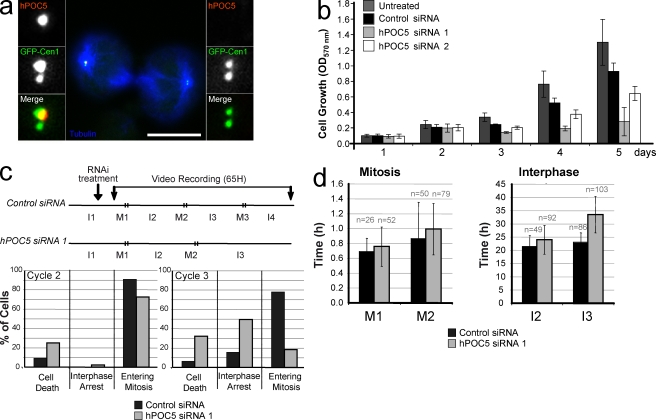
hPOC5 is an essential protein. (a) Dividing GFP-Cen1–expressing HeLa cells after 48-h treatment with hPOC5 siRNA 1 and stained with anti-hPOC5 (red) and anti-tubulin (blue). Insets show magnifications of each spindle pole area. (b) Cell growth is affected by hPOC5 depletion. HeLa cells were siRNA treated twice at t = 0 and 2 d later and fixed at designated time points, and cell growth was determined after violet crystal staining (OD570). Results were obtained from three independent measurements in three independent experiments. Mean values ± SD are shown. (c) Fate of HeLa cells treated with hPOC5 siRNA 1 observed by time-lapse imaging. (top) Experimental protocol. Asynchronous HeLa cells were treated with hPOC5 siRNA 1 or control siRNA and video recorded for 65 h starting 6 h after the beginning of siRNA treatment. I, interphase; M, mitosis. (bottom) Percentages of interphase cells that either died, arrested in interphase until the end of the recording, or entered mitosis during interphase 2 (left) or 3 (right). (d) hPOC5-depleted cells exhibit a delay in interphase. Duration of mitosis and interphase of HeLa cells treated with hPOC5 siRNA 1 or control siRNA. Results in c and d were obtained by analysis of time-lapse images of individual cells from two independent experiments. n = total number of cells counted. Error bars represent SD. Bar, 10 µm.

