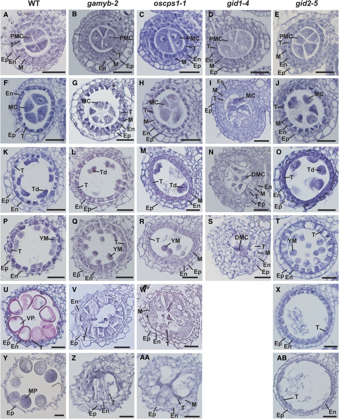
Figure 1.
Histological Analysis of Anther Development in Wild-Type and GA-Related Mutants.
Anther development in wild-type and GA-related mutants was compared at the six different stages: PMC, MEI, TD, YM, VP, and MP. Ep, epidermal cell layer; En, endothecial cell layer; M, middle layer; T, tapetal layer; MC, meiocyte; DMC, degraded meiocyte; Td, tetrad. Bars = 25 μm.
(A) to (E) Transverse sections of anthers at the PMC stage in the wild type (A), gamyb-2 (B), oscps1-1 (C), gid1-4 (D), and gid2-5 (E).
(F) to (J) Transverse sections of anthers at the MEI stage in wild type (F), gamyb-2 (G), oscps1-1 (H), gid1-4 (I), and gid2-5 (J).
(K) to (O) Transverse sections of anthers at the TD stage in the wild type (K), gamyb-2 (L), oscps1-1 (M), gid1-4 (N), and gid2-5 (O).
(P) to (T) Transverse sections of anthers at the YM stage in the wild type (P), gamyb-2 (Q), oscps1-1 (R), gid1-4 (S), and gid2-5 (T).
(U) to (X) Transverse sections of anthers at the VP stage in the wild type (U), gamyb-2 (V), oscps1-1 (W), and gid2-5 (X).
(Y) to (AB) Transverse sections of anthers at the MP stage in the wild type (Y), gamyb-2 (Z), oscps1-1 (AA), and gid2-5 (AB).

