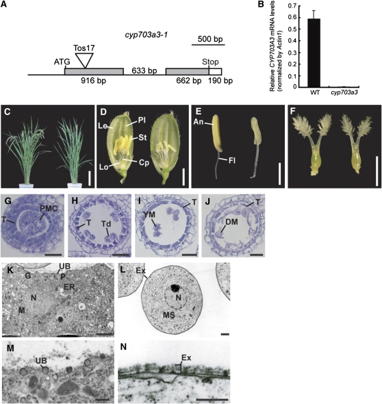
Figure 10.
Characterization of a Loss-of-Function Mutant of CYP703A3.
(A) Site of Tos17 insertion in the CYP703A3 gene. Untranscribed and intron regions are represented as lines. Coding and untranslated regions are represented as gray and open boxes, respectively. The insertion site of Tos17 is indicated as a triangle. Bar = 500 bp.
(B) Quantitative RT-PCR analysis of CYP703A3 in flowers. Total RNA was extracted from wild-type and cyp703a3-1 flowers. The transcript was assayed by real-time PCR relative to an internal Actin1 control. Data are means ± sd from three replicates.
(C) Gross morphology of the wild type (left) and the cyp703a3-1 mutant (right) at the ripening stage. Bar = 20 cm.
(D) Flowers of the wild type (left) and cyp703a3-1 (right). Cp, carpel; Le, lemma; Lo, lodicule; Pl, palea; St, stamen. Bar = 2 mm.
(E) Stamens of the wild type (left) and cyp703a3-1 (right). An, anther; Fl, filament. Bar = 1 mm.
(F) Pistils of the wild type (left) and cyp703a3-1 (right). Bar = 1 mm.
(G) to (J) Transverse sections of cyp703a3-1 anthers at the PMC stage (G), TD stage (H), YM stage (I), and MP stage (J). T, tapetal layer; Td, tetrad; DM, degraded microspore. Bars = 25 μm.
(K) to (N) Ultrastructural analysis of cyp703a3-1 anthers by TEM.
(K) The cytoplasm of tapetal cell in cyp703a3-1. N, nucleus; P, plastid; M, mitochondria; ER, endoplasmic reticulum; G, Golgi body; UB, Ubisch body. Bar = 1 μm.
(L) The cytoplasm of a cyp703a3-1 microspore. MS, microspore; N, nucleus; Ex, exine. Bar = 2 μm.
(M) Defective Ubisch body on the plasma membrane of tapetal cells in cyp703a3-1. Bar = 500 nm.
(N) Defective exine on the microspore coat in cyp703a3-1. Bar = 250 nm.

