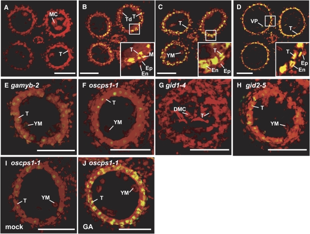
Figure 2.
TUNEL Assay of Anthers in Wild-Type and GA-Related Mutants.
(A) MEI stage in the wild type.
(B) TD stage in the wild type.
(C) and (E) to (H) YM stage in the wild type (C), gamyb-2 (E), oscps1-1 (F), gid1-4 (G), and gid2-5 (H).
(D) VP stage in the wild type.
(I) Mock-treated anther at the YM stage in oscps1-1.
(J) GA3-treated anther at the YM stage in oscps1-1.
The box in each panel outlines the portion of the anther cell layers shown at higher magnification in the inset. Nuclei were stained with propidium iodide (red), while yellow fluorescence is a TUNEL-positive signal. Ep, epidermal cell layer; En, endothecial cell layer; M, middle layer; T, tapetal layer; MC, meiocyte; DMC, degraded meiocyte; Td, tetrad. Bars = 50 μm.

