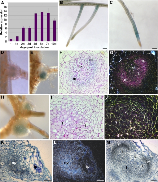
Figure 5.
Expression Analysis of S. rostrata CCaMK during LRB Nodule Development.
(A) qRT-PCR on developing stem nodules, relative to the constitutive S. rostrata UBI1. Tissues used are peels taken from the stem containing the dormant adventitious roots that were treated as described. Error bars indicate sd (n = 3).
(B) to (J) CCaMK:GUS expression in S. rostrata hydroponic roots. GUS staining is visible in uninoculated roots ([B] and [C]) and at 2 (D), 3 (E), and 6 (H) DAI with A. caulinodans ORS571. Microscopic sections are shown of 4-d-old ([F] and [G]) and 6-d-old ([I] and [J]) developing nodules viewed under bright-field and dark-field optics (signals seen as blue and pink spots, respectively).
(K) to (M) In situ transcript localization of CCaMK during stem nodule development at 3 ([K] and [L]) and 4 (M) DAI. In bright-field and dark-field images, signals are seen as black and white spots, respectively.
f, fixation zone; i, infection zone; ic, infection center; m, meristem; np, nodule primordium. Bars = 500 μm in (B) to (E) and (H) and 100 μm in (F), (G), and (I) to (M).

