Figure 2.

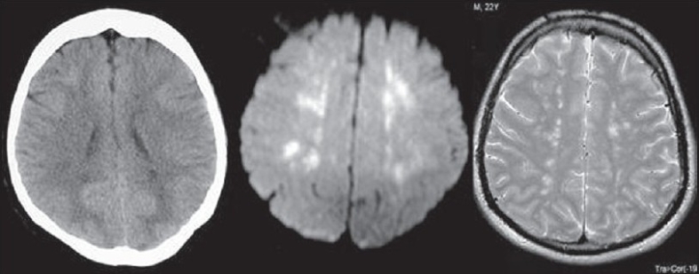
CT image showing minimal hypodense changes in periventricular region, which are more evident in DWI and T2WI as areas of high signals. Constellation of findings along with clinical data is characteristic for FES.

CT image showing minimal hypodense changes in periventricular region, which are more evident in DWI and T2WI as areas of high signals. Constellation of findings along with clinical data is characteristic for FES.