
Figure 4.
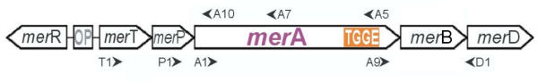
Schematic representation of a typical broad-spectrum mercury resistance operon of Gram-negative bacteria and the location of primers (arrowheads) used for amplification of merA. The merA stretch used for TGGE analysis is marked orange. R, regulator protein merR; OP, operator/promotor region and start of transcription; T, periplasmic transport protein merT; P, membrane bound transport protein merP; A, mercuric reductase merA; B, mercuric lyase merB; D, regulatory protein merD.