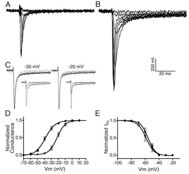
Figure 2.
ι-RXIA modulates the activity of NaV1.6 coexpressed with β1 in Xenopus oocytes by shifting its voltage-dependence of activation. Oocytes were two-electrode clamped at a holding potential of −100 mV, and Vm was stepped in 10 mV increments as described in Methods. Representative current records for voltage steps ranging from −60 to 10 mV in the absence (A) and presence (B) of 10 μM ι-RXIA. C, Normalized current traces from A and B in response to the voltage step to −30 mV (left) and −20 mV (right), in the absence (dots) and presence (solid line) of 10 μM ι-RXIA; each pair of responses to a given voltage step largely overlap. Insets show corresponding traces non-normalized. Voltage sensitivity of activation (D) and inactivation (E) obtained in the absence (open circles) and presence (closed circles) of ι-RXIA. The solid lines are best-fit curves to the Boltzmann equation. V1/2 values for activation in D were −22.38 ± 0.48 (control) and −38.29 ± 0.47 mV (10 μM ι-RXIA) with respective slope factors of −6.08 ± 0.42 and −7.53 ± 0.41 mV. V1/2 values for inactivation in E were −58.71 ± 1.03 (control) and −55.73 ± 0.75 mV (5 μM ι-RXIA) with respective slope factors of 5.98 ± 0.90 and 5.37 ± 0.61 mV. Each data point represents mean ± SE (N = 3 oocytes).

