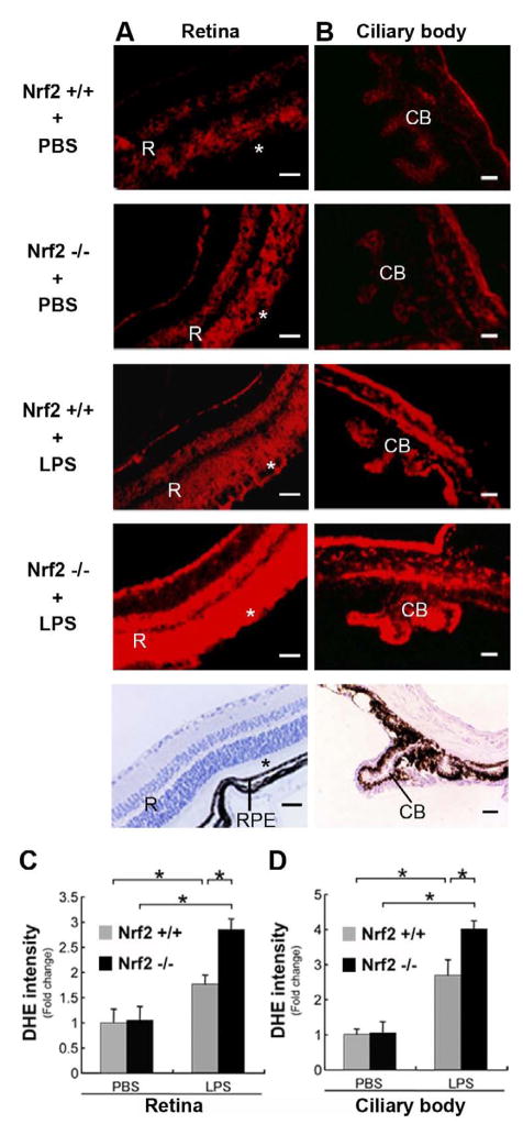
Figure 1.
Evidence of ROS in the Neurosensory Retina and Ciliary Body. DHE histochemical staining of ROS. A. Neurosensory retina of wild-type (Nrf2+/+; n=4) and Nrf2 deficient (Nrf2−/−; n=4) mouse shows minimal red label in the retina (R), photoreceptor layer of the retina (*), and retinal pigmented epithelium (RPE) when unstimulated (PBS treated). Increased DHE labeling in the retina of Nrf2+/+ (n=4) and Nrf2−/− (n=4) mice after LPS stimulation. Note marked increase in Nrf2−/− mouse retina. B Ciliary body processes of Nrf2+/+ and Nrf2−/− mice showed increase in DHE labeling after LPS treatment compared with PBS treatment. Note marked increase in Nrf2−/− mouse after LPS stimulation. The lowest panels show the hematoxylin staining in Nrf2+/+ mice. Bar = 100μm. (C, D) The graphs show the ratio of DHE fluorescent intensity to PBS-treated Nrf2+/+ mice. *p<0.05.

