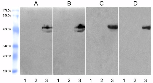
Figure 4.
Characterization of molecular weight of sCD226. Sera from healthy individuals (A) and cancer patients (B), or supernatant from PMA-activated PBMC (C) and PMA-activated Jurkat cells (D) were precipitated and detected as following: Lane 1, the samples were precipitated with normal mouse IgG-Sepharose 4B (negative control for immunoprecipitation) and detected with FMU4 (anti-CD226 mAb) in Western blot. There was no band because sCD226 could not be precipitated by normal mouse IgG-Sepharose 4B. Lane 2, the samples were precipitated with anti-CD226 mAb, LeoA1-Sepharose 4B and detected with anti-SED (staphylococcal enterotoxin D) mAb (negative control for blotting reagent) in Western blot. There was still no band because the precipitated sCD226 could not be detected with anti-SED mAb in Western blot. Lane 3, the samples were precipitated with LeoA1-Sepharose 4B and detected with the other anti-CD226 mAb, FMU4, in Western blot. There were bands because sCD226 could be precipitated by LeoA1-Sepharose 4B and detected with FMU4 in Western blot. One representative experiment is shown (n = 3).

