
Figure 1.
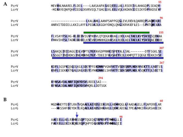
Sequence alignment between PcrV (P. aeruginosa) and LcrV (Y. pseudotuberculosis), and PcrG and LcrG. Identical residues are highlighted, and the predicted coiled coil region in PcrV is identified with a green bar. The blue bar represents the major antigenic region of LcrV identified in active and passive immunization studies [12]. The arrow points to the end of the PcrG (1–74) construct.