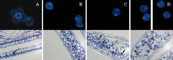
Fig. 1.
DAPI staining of barley isolated microspores (top) and semithin sections of anthers stained with toluidine blue (bottom). a Anthers before mannitol treatment. b, c, d Anthers after 4 days of mannitol stress treatment of DH46 (b), DH6188 (c), and DH6004 (d). (T tapetum, Black arrows indicate binucleate microspores)

