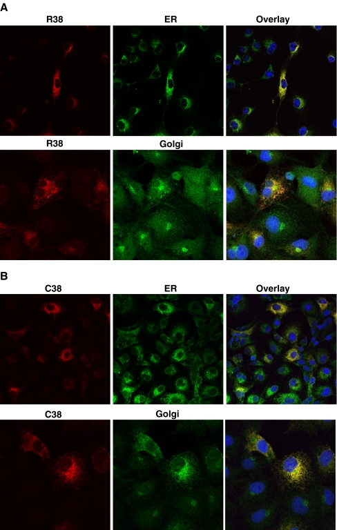
Fig. 3.
Subcellular immunofluorescence localization of hGX sPLA2 in COS-7 cells transiently transfected with vectors encoding either (a) R38 or (b) C38 hGX sPLA2. Twenty hours after transfection cells were fixed and stained with rabbit polyclonal against hGX sPLA2 (red) and either a mouse monoclonal against the ER marker PDI (green), or a mouse monoclonal against the Golgi apparatus marker 58 K protein (green). Nucleus is labelled in blue with DAPI Confocal analysis was performed on a Leica SP2-AOBS confocal microscope as described in “Materials and methods”. Magnification 50 for ER and 100 for Golgi

