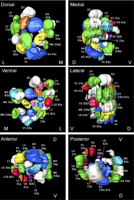
Fig. 3.
OSN representation in calyx glomeruli. This is a 3D reconstruction of a single calyx, viewed from different angles. GH146-expressing glomeruli are color-coded according to predicted input as in Fig. 1E; other glomeruli are white. Labeling indicates both the name of the best-fitting calyx glomerulus and the AL glomerulus connected to it. D, V, M, and L denote dorsal, ventral, medial, and lateral faces of the calyx, respectively.

